Coronavirus can cut cardiac fibroblasts into pieces
A new study discovers, coronavirus appears to cut heart fibre into tiny, precise pieces -- at least when it infuses heart cells in a lab dish.
Cutting muscle fibres into small pieces can permanently damage heart cells, as shocking as it is in a lab dish. But researchers have found evidence that a similar process is taking place in the hearts of patients with COVID-19. However, this new findings have only been published on 25 August in the bioRXiv preprint database, and have not been published in peer-reviewed journals or confirmed in real life.
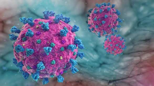
The findings were different from what the researchers had seen before-no disease affected heart cells in this way. “What we observed was absolutely not normal,” Todd McDevitt, co-author of the study, said in a statement.
This new finding could explain how COVID-19 can damage the heart. Previous studies have found abnormal signs of heart in COVID-19 patients, including inflammation of the heart muscle, even in mild cases.
For the new study, the researchers used special stem cells to create three types of heart cells -- cardiac myocytes, cardiac fibroblasts, and endothelial cells. Three heart cells in the petri dish were then exposed to SARS-CoV-2 virus. In all three types of cells, the virus can only infect cardiomyocytes and duplicate itself.
Cardiomyocytes contain muscle fibres that make up the myofibril, which is essential for the contraction of muscles to produce a heartbeat. These myofibril segments usually line up in one direction to form long fibres. But lab dish studies have revealed something strange - these myofibroblasts are cut into small pieces.
Study co-author Doctor Bruce Conklin stated, “the fragmentation of myofibril segments that we found in lab dish can cause heart muscle cells to fail to beat correctly.”
But findings in lab dishes do not necessarily apply to real-life medical science. So, the researchers analysed heart tissue samples from three new patients. They saw that myofibril fibres were disorganized and rearranged -- a similar pattern that seen in lab dishes, but not identical.
More research is needed to determine whether the changes in the myofibril segments of heart cells are permanent. The authors point out that scientists need to take a special, infrequently used method of looking at the myofibril segments to understand why this finding has been ignored in autopsies so far.
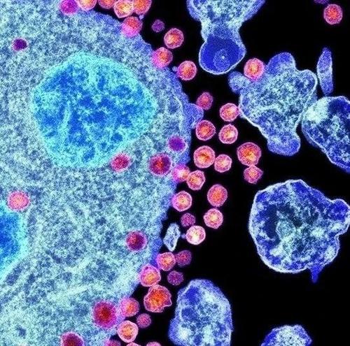
McDevitt said, “hopefully our results will inspire doctors to look back at their own patient samples and look for these features.”
The researchers observed another strange matter in both lab dish tests and in the heart tissue of patients with COVID-19. They discovered that in some heart cells, the DNA in their nuclei seemed missing. The authors say, this leaves the cells “brain dead” and unable to perform their normal functions.
Once scientists understand the mechanism by which form that Coronavirus destroys heart cells, they will be able to screen for specific drugs. For example, if the virus makes an enzyme to cut the myofibril, it might be possible to find a drug to interrupt the enzyme. (However, the authors indicate that it is unclear whether Coronavirus will directly cut the myofibrils or get cells cut the fibres by other mechanism)
“It’s important to find a protective treatment, a method to protect the heart from the sort of damage we see in the model,” McDevitt said. ”although we can’t stop the virus infecting the cells, we can give patients drugs to prevent these negative effects.”
OTHER NEWS
-
- Acer Global Launch Event, 2020
- By Johnny 24 Apr,2023

-
- Latest Report on Digital Payment in Southeast Asia
- By Marilyn 24 Apr,2023

-
- Video Mentoring Platform Superpeer Launches Paid Channels
- By Judy 24 Apr,2023

-
- A Black Friday With Booming Online Sales
- By Hughes 24 Apr,2023

-
- Zuckerberg Criticized Apple for Its Privacy Policy, Seeing Apple as One of Facebook’s “Biggest Competitors”
- By Anderson 24 Apr,2023

-
- Google Maps to Show the Locations of Traffic Lights
- By Jackson 24 Apr,2023

-
- How to Get Free Gold Bars in Candy Crush?
- By Davis 24 Apr,2023

-
- British scientists recommend people to add extra vitamin D to milk and bread
- By Betty 24 Apr,2023
-
- Cash: from the Most Popular Payment Method to the Least-used One
- By Shawn 24 Apr,2023

-
- Genetic factors have significant impacts on the risk of sex crimes
- By Paula 24 Apr,2023
-
- Playing video games as a child is proven to bring lasting improvement in cognitive ability
- By Jacqueline 24 Apr,2023
-
- How to Get Gold and Diamond for Free
- By Denise 24 Apr,2023

 1
1 1
1